Experimental Design:
4 month old wild-type mice were subjected to whole body irradiation, and then infused with bone marrow cells extracted from CAG-GFP mice, to generate bone-marrow chimeric mice. 3.5 months later mice were treated with the CSF1R inhibitor PLX3397 (600 ppm in chow), or vehicle, for 14 days, to deplete the microglial tissue. Withdrawal of the drug results in repopulation, which derives almost entirely from bone-marrow cells, effectively fully replacing the microglial tissue with infiltrating monocytes. See schematic below:
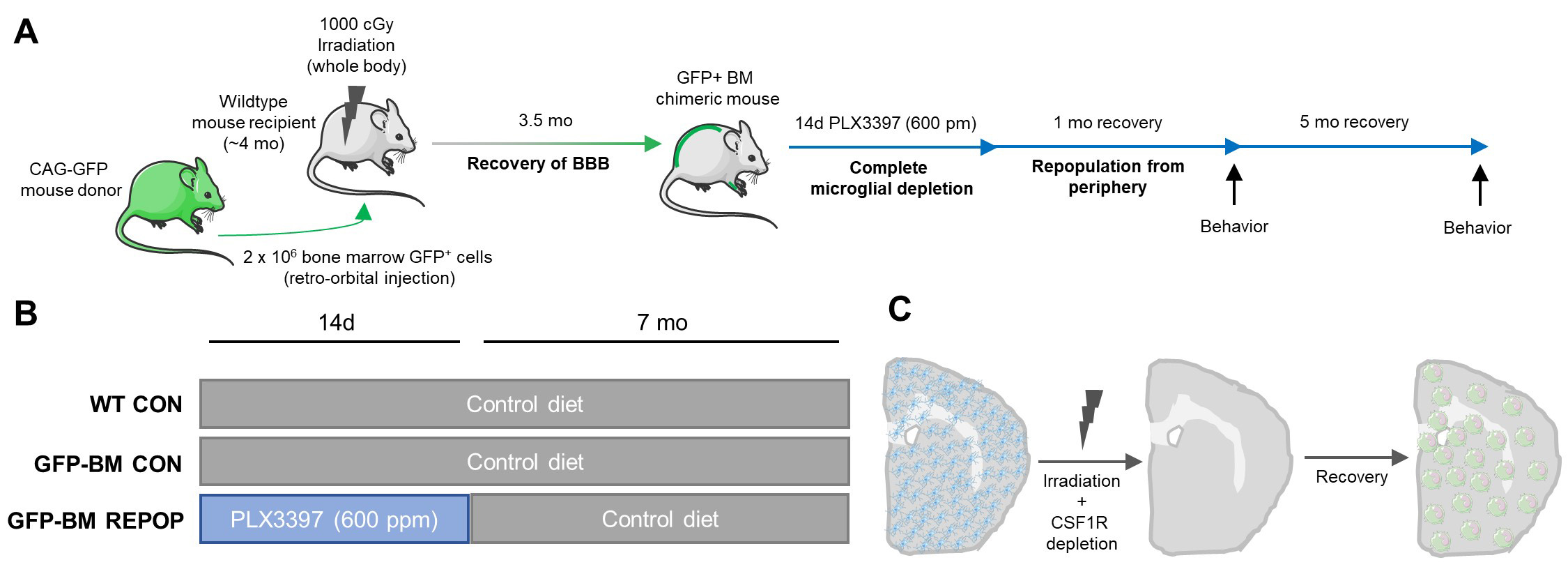
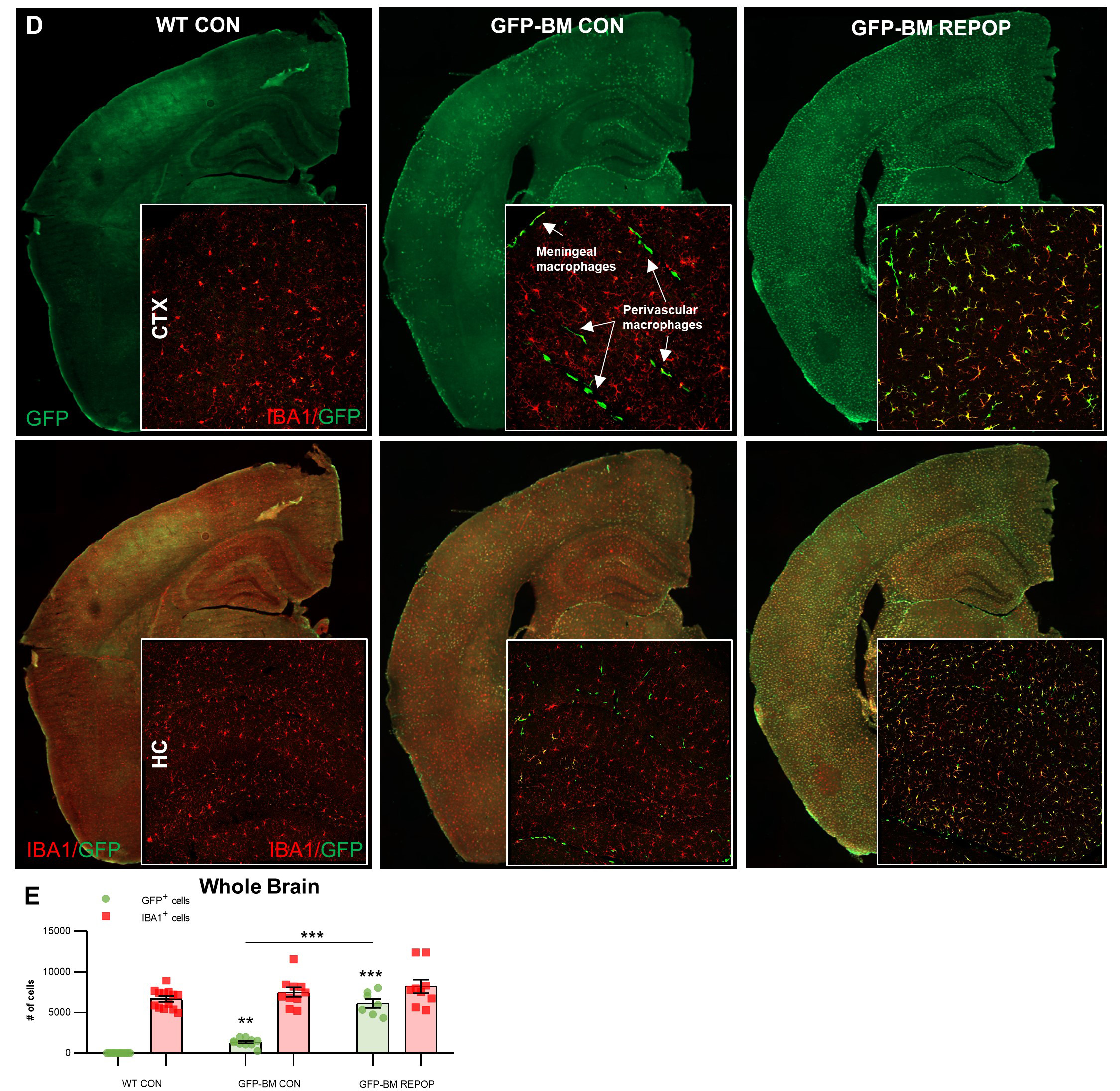
To explore the consequences of long-term monocyte engraftment in the brain, mice were then aged a further 6 months. Immunohistochemistry showed almost complete replacement of the microglial tissue with bone-marrow derived cells (shown as expressing GFP below). Irradiated controls were also included (recieved irradiation and CAG-GFP bone marrow transplant, but no PLX3397 to deplete and then replace the microglial tissue). Irradiated controls showed reconstitution of the perivascular and meningeal macrophages with bone marrow cells, but limited parenchymal monocyte infiltration. Brains were then microdissected into cortex, hippocampus, and thalamus/striatal fractions, RNA extracted, and sequencing performed.
Use the search box to the left to display raw expression data for any gene, expressed as RPKM (Reads Per Kilobase of transcript per Million mapped reads). Individual data points, as well as means and standard errors are displayed.
This dataset accompanies the manuscript "Selective vulnerability of the hippocampus to long term monocyte engraftment".