Experimental Design:
5xfAD and Wild-type mice treated with vehicle or PLX5622 (1200 ppm in chow) from 1.5 to 7 months of age. Brains microdissected into cortex, hippocampus, and thalamus+striatum. 4 mice/group.
This dataset allows for exploration of the effects of Alzheimer's disease pathology on disease expression in these 3 brain regions.
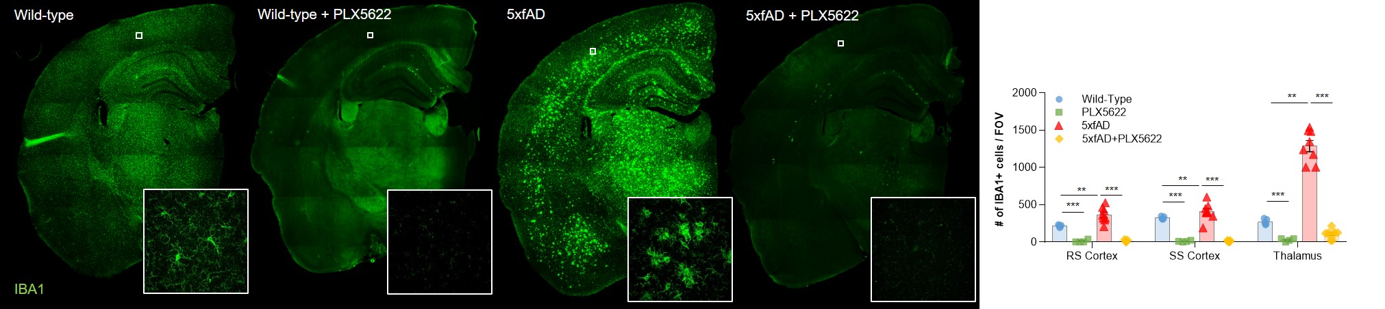
PLX5622 eliminates >95% of microglia in less than 5 days. Thus, these data also allow the exploration of the effects, and specificity, of 6 months microglial depletion on gene expression in these 3 brain regions, in both wild-type and 5xfAD mice. The figure below shows the extent of microglial depletion, with representative images (microglia identfied via IBA1 immunostaining), and quantfications shown on the right.
Use the search box to the left to display raw expression data for any gene, expressed as RPKM (Reads Per Kilobase of transcript per Million mapped reads). Individual data points, as well as means and standard errors are displayed.
This dataset accompanies the manuscript "Synthesis of a specific CSF1R inhibitor for sustained microglial depletion reveals crucial roles of microglia in plaque development and transcriptional alterations in Alzheimer’s disease mice".